
Neurosurgical Procedure Helps Some Forms of Cerebral Palsy
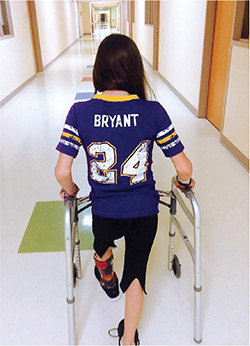 Marina Pellicciari, a Kobe Bryant fan, met the basketball star when her family took her to California in February for a Los Angeles Lakers game.
Tween Marina Pellicciari is a force on the basketball court. But she has cerebral palsy, and the muscle spasticity that often affects people with cerebral palsy was causing her pain and hampering her game.
Nearly 80 percent of children with cerebral palsy have spasticity, an involuntary stretching reflex that stiffens muscles, making it difficult to coordinate movement. Children with lower-limb spasticity usually walk on their toes, knees pointed inward.
“Kids with spastic CP spend significantly more energy coordinating their movement, which results in pain, fatigue, and postural problems,” says Richard Anderson, MD, associate professor of neurological surgery at CUMC.
Treatments range from intensive physical therapy to an implantable pump that delivers a steady dose of muscle relaxant, but most treatments do not address the underlying cause of spasticity. One procedure—selective dorsal rhizotomy, or SDR—gets to the root or, rather, the nerve rootlets of the problem. In SDR, the neurosurgeon cuts tiny, spaghetti-like strands of sensory nerve fibers at the base of the spinal cord. These nerve rootlets normally prevent muscles from overreacting to signals to contract, but the fibers are damaged in cerebral palsy, leading to muscle stiffness and spasticity.
Not everyone with spasticity is a candidate for SDR; it is only effective when spasticity is the main feature of a patient’s condition, before orthopedic complications have manifested. And recovery can take up to a year.
“Marina was a great candidate because she had significant spasticity and pain that were not adequately addressed with other therapies,” says Dr. Anderson. “Plus, her positive mindset would allow her to get through the postop rehabilitation required to learn how to walk again in a more physiologic way.”
During SDR procedures, Dr. Anderson uses a technique, currently performed at only a few medical centers in the United States, to minimize the amount of bone removed to reach the nerve roots. This reduces pain, recovery time, and the chance of spinal instability after the procedure.
The surgeon electrically distinguishes between motor and sensory nerve roots, then separates the individual sensory rootlets and applies mild electrical stimulation to each one.
“Less current is required to activate a dramatic response in motor nerves compared with sensory nerves, so we are able to guide the surgeon toward the sensory rootlets, and away from motor rootlets, with great confidence,” says Edward Gallo, technical supervisor of clinical neurophysiology at Columbia’s Comprehensive Epilepsy Center.
Patients treated with SDR have significantly better outcomes in terms of reduced spasticity, improved range of motion in the joints, and overall function compared with patients receiving an implantable pump, according to a large retrospective matched cohort trial. SDR patients also need less orthopedic surgery than pump patients.
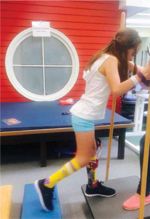
Marina had the surgery when she was 11. In her case, her functional abilities surpassed her baseline after just four months, including the seven weeks she spent in intensive rehabilitation to learn how to walk again. She is back to shooting hoops.
“Marina’s recovery and progress have been exceptional,” says Dr. Anderson. “I think this shows that considering a patient’s positive attitude is important when we select the ideal candidate for this procedure.”
For more information about SDR and spasticity management in children, contact Richard Anderson, MD, at 212-305-0219 or via email, rca24@columbia.edu.
- Log in to post comments