

More Than Skin Deep
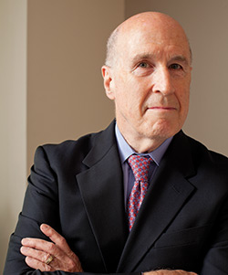 David Bickers, MD
When geneticist Angela Christiano showed histological slides from the skin of a patient with the hair-loss disease alopecia areata to immunologist Raphael Clynes, “He scratched his head and said, ‘Why is no one working on this?”
The pictures showed a swarm of T cells (lymphocytes) surrounding, and attacking, a hair follicle, a sure sign of the autoimmune disease. These basics of alopecia areata have been known for decades, but details that could lead to a treatment that restores hair growth were lacking. And few researchers were pursuing the leads.
“Alopecia areata can sometimes get pushed to the back burner,” Dr. Christiano says. “It’s not life-threatening, so it’s believed that it’s not a big problem. It’s just hair.”
It is true that no one dies from alopecia areata, but it is a life-altering disease, Dr. Christiano emphasizes with an unusual air of authority for a research scientist. She is also a patient. Soon after joining the Department of Dermatology in 1996, “my hair started falling out in patches typical of alopecia areata,” she recalls. The next two years were emotionally draining. Her hair eventually grew back, but it gave her a new research direction. Finding new ways to treat alopecia areata and other hair loss conditions became her lab’s major focus.
Dr. Christiano had reached a turning point in this search in 2010, after trawling the genomes of a thousand alopecia areata patients for new clues to the source of the disease. “We found a surprising overlap in genes linked to alopecia areata and autoimmune diseases such as type 1 diabetes, celiac disease, and rheumatoid arthritis.” Armed with these data and the pictures, she approached Dr. Clynes for advice.
“There’s a joke among geneticists that you can’t get an immunologist to return an email much less to talk to you,” says Dr. Christiano. Naomi Berrie Diabetes Center researcher Domenico Accili recommended that Dr. Christiano get in touch with Dr. Clynes, an expert in the study of cytotoxic (killer) T cells in diabetes.
“I remember the picture of a hair follicle swarmed by killer T cells, like bees attacking in a swarm—and the rest of the skin looked completely empty,” says Dr. Clynes. “I saw immediately that there was an elegance involved. It seemed like a simple problem to figure out, because it was confined to this one spot.”
Drs. Clynes and Christiano began collaborating and now, just four years later, they may be on the brink of delivering an effective drug treatment for the 6 million people in the United States with some form of the disease.
In a paper published in Nature Medicine in August of this year, the two reported results from the first three participants in a clinical trial directed by Julian Mackay-Wiggan, MD, assistant professor of dermatology and director of the department’s clinical research unit. The drug stops the T cell attack on the follicles, and in three patients with severe disease, nearly all hair returned after up to six months of therapy.
“This is a major step forward,” says David Bickers, MD, the Carl Truman Nelson Professor of Dermatology and chair of the Department of Dermatology at P&S. “There are few tools in the arsenal for the treatment of alopecia areata that have any demonstrated efficacy.”
The success in finding a potentially groundbreaking treatment for millions of alopecia areata patients clearly has much to do with Dr. Christiano’s personal quest, but it also is a testament to the collaboration between researchers from two separate fields, dermatology and immunology.
“People used to think that the skin was just a passive barrier, that it was simply there to protect the body from environmental stresses and there was nothing of interest going on,” says Dr. Bickers. “Now it’s very clearly a major outpost of the immune system, and it’s my belief that it is also the furthest outpost of the nervous system. The whole paradigm has changed and it’s why we’ve recruited researchers who can tap into the expertise of these other fields.”
The strategy of branching out into other fields has helped the department increase its share of NIH funding; it consistently ranks in the top 10 dermatology departments in the United States in terms of NIH funding. Since 1997 it has been designated as one of six Skin Disease Research Centers by the NIH, a coveted distinction that provides funding for interdisciplinary research projects. “The center allows us to create a variety of core facilities and fund pilot studies where people working in disciplines outside of dermatology can submit proposals that might connect to skin,” says Dr. Bickers.
And though the timeline of the Christiano and Clynes research—moving from genetic findings to positive results in a clinical trial in only four years—was astoundingly fast, says Dr. Bickers, it is just one example of how the department’s commitment to interdisciplinary discovery is paying dividends for patients.
Cyclops Sheep and a New Treatment For Skin Cancer
It is estimated that at any one time, a third of Americans are grappling with a skin disease, from diseases that cause simple burning or itching to life-threatening melanoma. In 2005, a report from the Society of Investigative Dermatology and the American Academy of Dermatology Association calculated the annual economic burden of skin diseases to be more than $37 billion, including lost productivity at work.
Dermatology chair David Bickers and a colleague found a drug that can dramatically shrink basal cell skin cancers and prevent the formation of new cancers.
Dr. Bickers came to Columbia in 1994 with a major interest in skin cancer projects, including research into basal cell nevus syndrome, a rare genetic disorder that causes multiple basal cell carcinomas in patients. Surgery has been the only option for these patients until recently, when Dr. Bickers and Ervin Epstein of Children’s Hospital Oakland Research Institute in California found that a drug called vismodegib can dramatically shrink basal cell skin cancers and prevent the formation of new ones. Their findings were published in 2012 in the New England Journal of Medicine.
The new treatment for basal cell nevus syndrome, also called Gorlin syndrome, springs from an unlikely location: Idaho’s mountain pastures. In the 1960s and 70s, sheep farmers began noticing that when their flocks grazed at higher altitudes, pregnant ewes would give birth to lambs with a single, central eye. The alarmed sheep farmers called in the U.S. Department of Agriculture, which launched an investigation. It took 11 years to discover that the ewes were grazing on the American corn lily, a mountain meadow plant that contains a chemical that disrupts normal brain and facial development, resulting in the deformities the farmers observed.
The P&S Department of Dermatology has one of the longest-funded Skin Disease Research Core Centers (SDRC) in the nation, one of only six centers supported since 1988 by the National Institute of Arthritis and Musculoskeletal and Skin Diseases (NIAMS).
NIAMS supports centers that provide shared facilities and services to groups of established, funded investigators who address scientific problems in skin biology and diseases. The centers are designed to improve efficiency, accelerate the pace of research, and ensure greater productivity.
Columbia’s SDRC has been funded since 1997. Its major goal is to build and sustain awareness of the opportunities that exist for discovering the pathologic basis of skin diseases that in turn can be translated into innovative treatments that will improve the quality of life for patients. The Columbia SDRC provides the infrastructure and support for ongoing research groups centered around three themes: skin neuroscience, stem cells, and genetics and immunology.
The department has been chaired by David R. Bickers, MD, since 1994. Before joining P&S, he was professor and chair of dermatology for 15 years at Case Western Reserve University, where he was awarded one of the two original SDRCs funded in 1988. The P&S Department of Dermatology consistently ranks among the best-funded dermatology departments in the nation. Since 2006 it has ranked in the top 10 in NIH research funding, including two years at the No. 2 position.
Years later, the corn lily chemical—cyclopamine—was determined to be a potent inhibitor of the Sonic hedgehog (Shh) signaling pathway that is critical for normal fetal development.
“I was also interested in looking at underlying mechanisms and trying to identify therapeutic targets in skin cancers, with the ultimate goal of finding drugs and compounds that could be taken to the clinic,” says Dr. Bickers. He knew that Gorlin syndrome patients inherit a mutation in a tumor suppressor gene family called patched (PTCH).
PTCH1 was the primary inhibitor of the Shh signaling pathway that was turned off in the cyclops sheep. Normally PTCH1 causes the signaling to cease after birth. But when PTCH1 is mutated, signaling continues, resulting in abnormal cell growth and proliferation and setting the stage for tumor formation. “If we could find a molecule to inhibit this pathway we could, in theory, reverse the growth of the tumors,” says Dr. Bickers.
The right molecule turned out to be cyclopamine, the corn lily chemical. Dr. Bickers, working with research scientist Mohammad Athar, PhD, and Arianna Kim, PhD, assistant professor of dermatology, found that while it had disastrous results for the farmers and their flocks, it was precisely what Gorlin syndrome sufferers needed: a way to block postnatal signaling that led to the unchecked growth of tumors in many patients.
Drs. Epstein and Bickers investigated the use of a vismodegib, a derivative of cyclopamine synthesized by Genentech. Their clinical study found that patients taking this orally administered drug for an average eight months experienced about two new basal cell cancers during the study, compared with 29 such tumors for patients in the placebo group. Among patients taking the drug, the diameter of clinically significant skin cancers decreased an average of 65 percent, compared with 11 percent among controls.
“The purpose of the study was to see whether, by using this targeted molecular therapy, we could match the performance of a surgeon, and in many ways, we could,” says Dr. Bickers.
Important issues remain, however, and Dr. Bickers says the drug is not suitable for all patients with basal cell nevus syndrome. The drug caused muscle pain and loss of taste in many patients, and half of the study’s participants stopped using the drug even though that meant their cancers returned.
“The challenge now is to see if we can lessen the adverse effects while achieving the same therapeutic benefits by modifying the dosing schedule or perhaps by alternating drug treatment with other modalities such as photodynamic therapy, which can be effective for smaller lesions.”
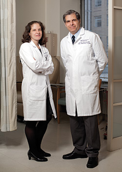 Yvonne Saenger, MD, and Gary Schwartz, MD
Melanoma Makeover
Seven years ago, patients with malignant melanoma had one treatment option: chemotherapy with dacarbazine, which had devastating side effects and did not prolong survival. The first major breakthrough was the identification that 50 percent of all melanomas carry an oncogene called B-RAF that is responsible for melanoma cell growth. This resulted in the development of two drugs (vemurafenib and dabrafenib) that inhibit this oncogene. These drugs show amazingly high response rates in this disease and in themselves result in a prolongation of survival in melanoma patients with advanced disease. “However, there are some limitations with this class of drugs in that patients often develop resistance to them and they essentially stop working,” says Gary Schwartz, MD, chief of hematology and oncology and associate director of the Herbert Irving Comprehensive Cancer Center. “In view of this there has been renewed interest in the immune system and trying to find ways to harness one’s own immunity as a means to treat melanoma. In particular, we asked if there was a way to turn on the immune system to target cancer cells and not the healthy cells of the body.”
Renewed interest in the immune system and trying to find ways to harness a person’s own immunity to treat melanoma has helped transform the disease from a mostly incurable one to one that is manageable for months to years in some patients.
Collectively, this work has helped transform the disease from one that was virtually incurable to one that is manageable for months to years in some patients. Dr. Schwartz and colleagues at Memorial Sloan Kettering, where Dr. Schwartz was chief of the melanoma and sarcoma service until moving to Columbia earlier this year, found that a drug called ipilimumab could restore the immune system’s ability to attack cancer. Cancer shuts down the immune attack via a protein, CTLA-4, on the surface of T cells.
“Ipilimumab blocks this protein and activates the T cell,” says Dr. Schwartz. “In clinical studies with melanoma patients, we are seeing durable complete remissions for the first time, sometimes for as long as five years.”
Because side effects with ipilimumab, which was approved by the FDA in 2012 and marketed as Yervoy, are still an issue, Dr. Schwartz and other researchers are now focusing on another T-cell protein, PD-1, a target that is associated with fewer side effects. “Five years ago, the two-year survival rate in patients with metastatic melanoma was 1 percent,” Dr. Schwartz says. “Now, our initial data shows that the two-year survival rate is approaching 90 percent. We have transformed metastatic melanoma from an incurable cancer to one that we think is now manageable—and perhaps curable—by harnessing the immune system.” To build Columbia’s immune targeted therapy program in melanoma, Yvonne Saenger, MD, an immuno-oncologist, was recently recruited from Mount Sinai School of Medicine to lead efforts to develop new therapeutic approaches directed at novel immune targets in melanoma. She also will focus her research studies on identifying specific biomarkers in tumors or in the blood by which she hopes to identify patients who will most benefit from this type of targeted therapy.
Looking toward the immune system also may lead to new therapies for another skin cancer: squamous cell carcinoma. Usually these common cancers can be easily treated with surgery, but in certain clinical settings these tumors are more aggressive and dangerous, killing about 2,300 people in the United States each year. Many of these deaths occur in patients who are immunosuppressed, such as organ transplant recipients. These patients require lifelong treatment with drugs that suppress the immune system to prevent rejection of the transplanted organ.
A basic researcher, David Owens, PhD, associate professor of epithelial cell biology (in dermatology, pathology & cell biology, and dental medicine), initially fell in love with skin as a model epithelial organ while he was a graduate student. “I got hooked when I started doing tumor experiments. In the skin you can see these tumors emerge right before your eyes,” he says. “I began wondering where the tumors come from.”
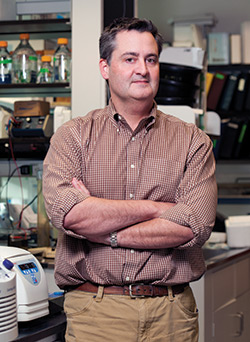 David Owens, PhD
Like melanoma, squamous cell carcinomas actively engage with immune cells to evade detection and fuel their expansion. “We think this interaction is critical to metastasis,” says Dr. Owens.
Using a mouse model, Dr. Owens has identified a signaling axis that may be essential for the ability of these cells to propagate secondary tumors. If so, the signaling axis could be utilized as a biomarker to indicate more aggressive cancers.
Working with Vishal Patel, MD, assistant professor of dermatology and a dermatologic surgeon who specializes in treating high-risk patients with skin cancer, Dr. Owens is using tissue samples collected in surgery in hopes the analyses could help him firmly establish the utility of this biomarker in identifying lesions that have a higher propensity to metastasize in humans, which could lead to treatments personalized to fit each patient’s needs.
“Our collaboration with Dr. Patel is designed to address two goals,” says Dr. Owens. “First, we hope to firmly establish the utility of this signaling axis as a biomarker that will allow us to identify cutaneous squamous cell carcinoma lesions that have a higher propensity to metastasize in humans, and, second, we want to identify ‘druggable’ tumor-specific targets of this axis as a novel therapeutic strategy for high-risk patients.”
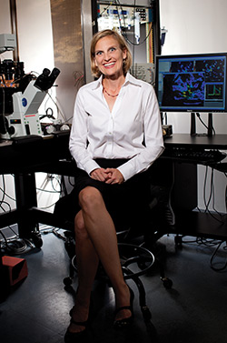
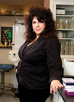 Angela M. Christiano, PhD
Stem Cell Biology
Before she turned her attention to alopecia, Dr. Christiano was on the hunt for genes that cause epidermolysis bullosa (EB), a rare disease of the skin and connective tissues that causes severe and even fatal blistering of the skin and mucous membranes. “As skin diseases go, epidermolysis bullosa is about as severe as it gets,” says Dr. Christiano.
As a postdoctoral student, Dr. Christiano worked in the lab that discovered the gene responsible for a specific subtype of the disorder. The mutation in the gene, COL7A1, causes a defect in type VII collagen that attaches the epidermis to the underlying dermis. Without this connection, the skin splits apart from even the lightest of touches.
After the discovery of COL7A1, epidermolysis bullosa became a target for conventional gene therapy. The idea was to take skin cells from a patient, correct the defective gene, and then graft the corrected skin back onto the patient.
“But there were a lot of challenges with that approach,” says Dr. Christiano. “There was no persistence of gene expression, and the grafts weren’t long term because they didn’t have stem cells, meaning that they could not self-renew.”
"This technology basically enables you to shake the Etch A Sketch and reprogram a patient’s own cells back to pluripotency so that you can then push them down a different pathway."
Dr. Christiano began to think about bone marrow transplants as a way to repair defective skin. “When patients have a bone marrow transplant, stem cells in the donated marrow repopulate the marrow in the recipient, but they also wind up in a lot of places, including the skin,” she explains. This observation led her to reason that if an EB patient were given bone marrow harvested from a person with normal COL7A1 genes, the stem cells would eventually find their way to the patient’s skin and other affected tissues and begin producing normal collagen.
Dr. Christiano suggested the transplant idea to pediatric transplant researchers at CUMC, led by Mitchell Cairo, MD, formerly in the Department of Pediatrics at P&S. Using Dr. Christiano’s preliminary data on an EB mouse model, the researchers were encouraged that the treatment was successful in animal tests before performing a transplant at CUMC on a patient using bone marrow cells from his unaffected brother.
Other centers around the world are also trying the approach, and now, close to 30 kids with EB have undergone the treatment. The down side: The transplanted patients require the same lifelong immunosuppressive drugs that other transplant recipients take.
Induced pluripotent stem (iPS) cells may be a better option in the future, says Dr. Christiano. “This technology basically enables you to shake the Etch A Sketch and reprogram a patient’s own cells back to pluripotency so that you can then push them down a different pathway. We saw the possibility of starting with cells from an EB patient, correcting the gene, and then grafting them back onto the patient.”
The discovery of iPS cells also shed new light on a well-known fact of
unrecognized significance: About one-third of patients with EB have healthy and intact areas of skin. “Previously doctors would largely ignore these patches in these patients,” says Dr. Christiano. “But it turns out that those patches have undergone a spontaneous correction event. That normal appearing skin is healthy and producing normal COL7A1. If we take those healthy cells
as starter cells for iPS, then we don’t have to go through the gene correction step. We can use the fact that the body has already corrected these cells and then we can generate unlimited healthy cells for use in treating these patients.”
Sense of Touch
Whenever Ellen A. Lumpkin, PhD, is at a party and guests find out she makes her living studying touch, “That’s all anyone wants to talk about,” she says. The sense of touch creates the sensation of a soft caress but is also necessary for more mundane tasks like manipulating pens and buttons.
In sensory neuroscience, says Dr. Lumpkin, associate professor of somatosensory biology (in dermatology and physiology & cellular biophysics), touch is the last frontier. The cells and molecules that initiate vision—rod and cone cells and light-sensitive receptors—have been known since the early 20th century, and the senses of smell, taste, and hearing are increasingly understood. But almost nothing is known about the cells and molecules responsible for initiating our sense of touch.
Dr. Lumpkin, who originally trained in the neuroscience of hearing, now focuses on understanding how the cells in skin transmit information about tactile sensations. She has solved a 100-year-old mystery about the nature of touch, which will let researchers start to ask even bigger questions.
For decades scientists have known that Merkel cells, specialized skin cells concentrated in fingertips, lips, and other highly sensitive areas, are involved in the sensation of gentle touch and pressure. The question was how: Are they simply scaffolding that allow touch signals to be transmitted or are they sensing and actively creating touch signals?
In two back-to-back studies published earlier this year in Nature, Dr. Lumpkin and colleagues showed that Merkel cells can sense touch and work virtually hand in glove with the skin’s neurons to create what we perceive as fine details and textures. Using optogenetics to turn the cells on and off, the researchers were able to identify the biophysical basis of touch sensitivity in Merkel cells.
 Ellen A. Lumpkin, PhD
“What’s more, we collaborated with Scripps scientists to pinpoint the gene that encodes the Merkel cells’ molecular touch sensors. These studies are the first to demonstrate that Merkel cells are capable of turning touch into an electrical signal that is then transmitted to the brain as the sensation of light touch.”
The knowledge of how nerve cells and Merkel cells divide up the job of creating and sending touch signals provides a jumping off point for touch researchers. “Now we can start to ask deeper questions, such as how the brain deciphers information sent by skin cells and nerves so that we can perceive shapes, textures, and pressure,” says Dr. Lumpkin.
The research also may lead to treatments for people who become touch-impaired, including people with diabetes and the elderly. “When we lose our sense of gentle touch, it has serious repercussions. In the elderly, this contributes to loss of handgrip and falling,” says Dr. Owens.
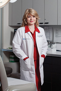
 Julian Mackay-Wiggan, MD
Merkel cells start disappearing from our skin in our 30s and 40s, and by the sixth or seventh decade, almost no Merkel cells are left. As a stem cell biologist, Dr. Owens became intrigued by the idea of restoring Merkel cells, which may be possible by reviving the right stem cells in the skin. “We know the stem cells are still there, but they are not making Merkel cells. We have to figure out how to entice them to reproduce.”
Clinical Research
When the findings from the Department of Dermatology’s basic researchers are ready to be tested in patients, the department’s in-house clinical research unit, directed by Dr. Mackay-Wiggan and including a research fellow and in-house research coordinator, speeds the process. “Being able to take that next step into translational and clinical research is vital,” says Dr. Christiano. “If we didn’t have a self-contained clinical trial unit in our department, our trials for alopecia areata would not have happened so quickly.”
Skin cancer patients at Columbia will see similar benefits with the department’s new comprehensive skin cancer center led by Larisa Geskin, MD, a renowned researcher in cutaneous T-cell lymphoma and associate professor of dermatology. Dr. Geskin’s dual training in dermatology and oncology allows her to build bridges between clinical disciplines and enables the center to support interdepartmental collaborations, such as the one between Dr. Owens and Dr. Patel. The center provides a place that brings together experts from across the medical center to consult on patients with skin cancer.
 Larisa Geskin, MD
“It is my philosophy that when you are a relatively small department, your success hinges on building collaborations and talent across the institution,” says Dr. Bickers. “We may be small, but we’ve been very fortunate to recruit and retain talented faculty and to find splendid research collaborators in other departments as well. Fortunately, Columbia is a place where such cooperative opportunities are a part of our culture.”
- Log in to post comments